| List of picture series | Menu | Lijst met fotoseries |
| 6. Digestion | All
pictures: / Alle foto's: © A.A. Verveen X-ray: / Röntgenfoto: RW.F. Becking, veterinarian |
6. Vertering |
| The dead rabbit which is situated in the stomach and esophagus of the female is quite well visible shortly after mealtime. |  |
Kort na het verzwelgen van het dode konijn zie je het nog goed, met het achterlijf ervan in de slokdarm kort voor de maag. |
| At
left a boa immediately before (L) and shortly after its meal (R). At right another boa shortly after its meal (L) and afyter digestion has been finished and she has emptied her bowels (R). |
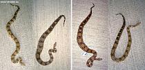 |
Links
zie je een boa direct voor (L) en direct na (R) de maaltijd. Rechts een boa direct na de maaltijd (L) en na afloop van de vertering als er ontlasting is afgezet (R). |
| Distension
of the boa occurs several days after the meal. It is caused by the formation of gases in the process of digestion of the prey. |
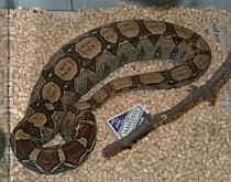 |
Enkele dagen na de maaltijd is de boa door bij het verteren van de prooi vrijkomende gassen flink opgezwollen. |
| Another example of the balloon-like gaseous expansion. | 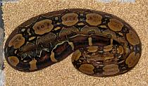 |
Nog een voorbeeld van het als een ballon opgeblazen zijn van de boa. |
| Pictured
time course of the process of expansion during digestion. Compare the
diameters that are posited on the white line. The boa has swallowed one mouse only. |
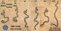 |
Fotoserie
van de uitzetting ten gevolge van de vertering van de prooi. Vergelijk
de doorsneden die op de witte streep liggen. De boa heeft één muis op. |
| Similar,
but after a meal of two mice. Note the feces deposited on the last day (August 25th). |
 |
Idem,
na een maaltijd van twee muizen. N.B. Let op de ontlasting die op de laatste dag (25 augustus) is afgezet. |
| After some days the snake needs to get rid of urates. It drinks water to be able to deposit urin.e | Enkele dagen na de maaltijd moet de boa de uraten kwijt. Het drinkt om die straks met de urine te lozen. | |
| Detail. | 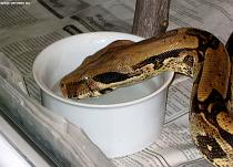 |
Detail. |
| Sequence to show the regular occurrence of the tongue to taste the water. Only the tip of the tongue is used in this situation. |  |
Serie opnames om het regelmatige proeven tijdens het drinken te laten zien. Alleen het uiterste puntje van de tong wordt hiervoor uitgestoken. |
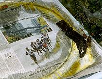
| Urine
deposit, here together with the faeces. The "solid" part is white, the
fluid discharge is yellow-brown. It stinks. |
 |
Urine
afzetting, hier plus de feces. De "vaste" urine is wit van kleur, de
vloeibare geelbruin. Alles stinkt. |
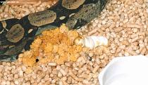
| Cat
litter removes the stink. Discharge of fluid as well as deposit of solid (white) blob of urine. |
 |
Kattenbakkorrels
nemen de geur goed weg. Afzetting van vaste (wit) en vloeibare urine. |
| The solid deposit may be coated with a yellow substance. | 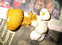 |
De vaste afzetting is soms omhuld door een stevige gele substantie. |
| Sometimes even of a nice orange colour. | 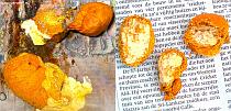 |
Soms ook fraai oranje. |
| A
solid deposit may also be coloured a yellow-brown throughout. The black substance is faecal. |
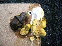 |
Soms
is de substantie geelbruin van kleur. De zwarte substantie is ontlasting. |
| A brownish cast of a ureter. | 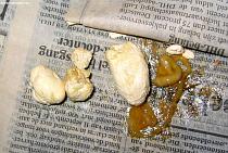 |
Een bruin afgietsel van de urinewegen. |
| Some
bones or teeth may escape digestion. X-ray taken 85 days after mealtime of a pregnant female boa, taken because her condition was in question. Vertebrae and ribs are white. Bones of the prey were tinted with yellow, gas bubbles with red. |
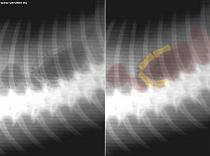 |
Sommige
tanden of botten ontsnappen aan de vertering. Rontgenfoto van een drachtige boa, 85 dagen na haar laatste maaltijd gemaakt omdat niet duidelijk was wat zij mankeerde. Ruggewervels en ribben zijn lichtgekleurd, botjes van de prooi zijn geel en gasbellen rood getint. |
| Hairs
of the prey are not digested and they stay in the stomach until
the very end of digestion after which they are quickly transported to
form the main ingredient of
the faeces. Anatomical position of the stomach (centre and top) relative to the length of the boa and with regard to the tip of the snout. |
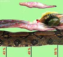 |
De
haren van de prooi verteren niet en blijven tot het laatste moment
in de maag, waarna zij snel worden doorgesluisd en het hoofdbestanddeel
van de ontlasting vormen. Anatomische positie van de maag (midden en boven), relatief ten opzichte van de lengte, gerekend vanaf de neuspunt. |
| Colon
of
a female that died 223 days after her last meal. Her death was due to an obstruction of the end of the ileum (right). Faecal remains are still present. |
 |
Opengeknipte dikkedarm van een vrouwtje dat 223 dagen na haar laatste maaltijd aan een darmafsluiting aan het eind van de dunne darm (rechts) stierf. Resten ontlasting zijn nog in de dikke darm te zien. |
| Defecation
usually occurs together with urinary discharge. At such a moment the true size of the tail is evident. |
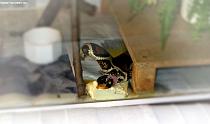 |
Bij
het ontlasten wordt meestal ook urine afgezet. Daarbij is dan duidelijk te zien hoe kort de eigenlijke staart is. |
| Usual shape of the stools. | 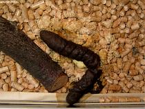 |
Normale ontlasting. |
| Dried
faeces, mainly composed of hair of the prey. Some of the rat's whiskers are clearly visible. |
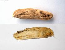 |
Ingedroogde
ontlasting. Deze bestaat voornamenlijk uit haren van de prooi. Enkele snorharen van de rat zijn zichtbaar. |
| Picked
dried out faeces. Mainly hair and (centre) bacteria. Some nails, teeth and bones in addition (see below). |
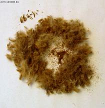 |
Uitgeplozen
ingedroogde ontlasting. eze bestaat hoofdzakelijk uit haren en bacterieën (centrum). Daarbij komen nog enkele botjes, nagels en tanden (hieronder). |
| Detailed
view. Nails, plus some teeth and bones of the rat. |
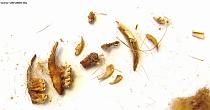 |
Detail. Nagels plus enkele tanden en botjes van de rat. |
| Revised: January 24th, 2008 | ||
| List of picture series | Top / Naar boven | Lijst met fotoseries |